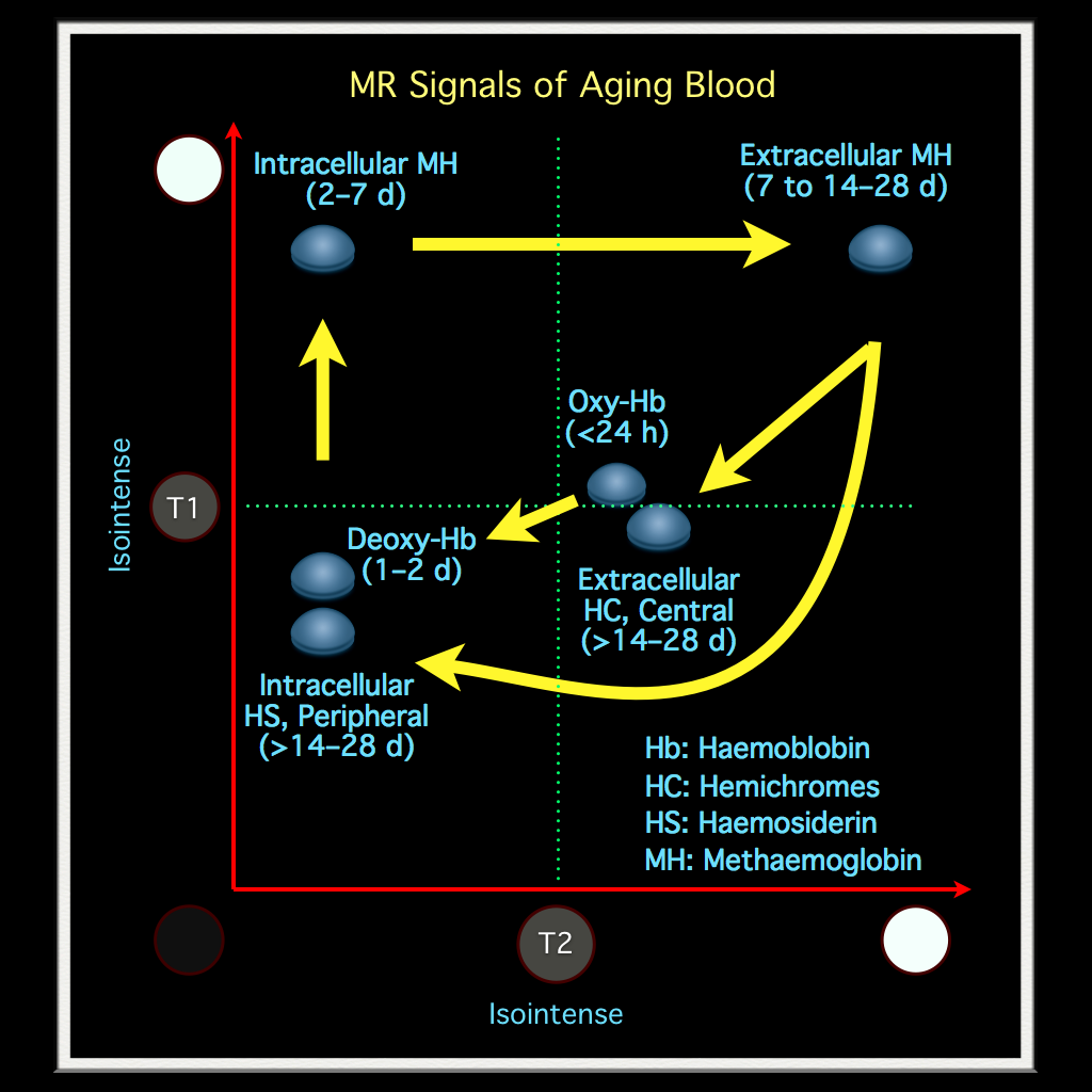
| Time | T1 | T2 | |
|---|---|---|---|
| Hyperacute | < 12 hr |
Isointense |
Bright |
| Acute | 12 hr - 2 days |
Isointense |
Dark |
| Early subacute | 2 - 7 days |
Bright |
Dark |
| Late subacute | 1 wks - 2 mos |
Bright |
Bright |
| Chronic | > 2 mos |
Dark |
Dark |
MRI Basic
Intensity Pattern
Mono
Compare intensity to grey matter.
T1
Hypo T1: fluid (chronic lesion)
Hyper T1:
- Fat (except
T1FS) - Protein (depend on content)
- Blood (early & late subacute)
- Paramagnetic, Melanin
- Fat (except
T2
- Hyper T2: fluid (CSF) > fat
Combined
Hypo T1 & T2: Air, Flow void (high-flow vessel), Calcification
Iso T1 & T2: Muscle
Hypo T1 + Hyper T2: fluid
Blood Signal
References
[1]
W.G. Bradley, MR appearance of hemorrhage in the brain, Radiology. 189 (1993) 15–26. https://doi.org/10.1148/radiology.189.1.8372185.
